Defining CT Dose Optimization Targets in Clinical Practice
January 12, 2026
Ensuring patient safety and consistent imaging quality remains a core priority in radiology departments. The International Atomic Energy Agency (IAEA) outlines clear goals for patient dose monitoring in its Safety Report Series 112 [1], emphasizing optimized radiation protection, imaging safety and accuracy of individual patients, justification and appropriateness, and awareness of collective dose from sources of medical exposure, among others. To support these aims, a study was carried out in the Ortenau-Klinikum Offenburg-Kehl in Offenburg, Germany, focusing on the implementation of the IAEA targets in clinical practice.
A state-of-the-art dose management system (DOSE, Qaelum, Belgium) was deployed across the five clinic locations. Variability was first assessed across two CT scanners (Somatom Definition AS+, Siemens, Germany) and two high-volume protocols: head with contrast (n=4617) and dual-phase abdomen (n=1709), performed throughout 2024. Radiographers documented reasons for increased dose values according to StrlSchV Anlage 14 (§108) [2] using the “Activity stream” feature of DOSE (Figure 1), which were then reviewed by radiologists to ensure transparent and consistent justification.

Figure 1. Activity stream feature of DOSE, allowing radiographers to indicate the cause of high dose exams, with further options to document the Action and Justification, when necessary.
Comparisons revealed strong alignment between scanners: standard study composition and scan settings were identical, while median CTDIvol and scan length differed by only 1% and 3%, respectively. However, a notable exception emerged during the 4 a.m. shift on one scanner, where CTDIvol exceeded the average by 20% (Figure 2). Contributing factors included use of higher kVp, patient body habitus, and suboptimal arm positioning.
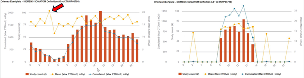
Figure 2. Hourly trend in mean and cumulative CTDIvol for the abdomen protocol on scanner 1 (left) and 2 (right). A 20% higher mean CTDIvol is observed on scanner 1 during the 4am shift (red arrow).
The dose management system enabled a detailed review of both low-dose and high-dose outliers using its outlier analysis tool. Most head studies falling below the 2nd percentile involved female patients, likely reflecting smaller head size, while abdomen studies often contained repeated localizers, suggesting potential challenges in positioning or protocol selection. At the upper end, studies exceeding the 98th percentile were linked to additional perfusion imaging (head) or large patient size (abdomen).
Using interquartile-range-based thresholds, 75 mild and 11 extreme outliers were identified within abdomen examinations. These were most often associated with high BMI, inappropriate kVp use, or scanning with arms down, reinforcing the importance of proper technique and protocol adherence.
Typical effective doses ranged from 1.9 to 27 mSv per study, though the system also highlighted examinations exceeding 100 mSv, particularly in abdomen with contrast and thoracic oncology imaging. Identifying and reviewing such cases is fundamental for strengthening justification workflows.
This project underscored several important opportunities for further optimization:
- Reducing repeated abdomen localizers through improved patient positioning.
- Reviewing image quality in low-dose outliers to ensure adequacy.
- Validating correct use of scan parameters across shifts.
- Enhancing data completeness, including operator initials and slice data.
This study was presented at the Annual Meeting of the German Society for Medical Physics (DGMP) in September, 2025 in Berlin [3].
If you want to know more about DOSE by Qaelum visit qaelum.com/solutions/dose or send an email to
References:
[1] INTERNATIONAL ATOMIC ENERGY AGENCY, Patient Radiation Exposure Monitoring in Medical Imaging, Safety Reports Series No. 112, IAEA, Vienna (2023).
[2] DEUTSCHE BUNDESREGIERUNG, Verordnung zum Schutz vor Schäden durch ionisierende Strahlung (Strahlenschutzverordnung – StrlSchV), insbesondere § 108 i. V. m. Anlage 14, Bundesgesetzblatt Teil I Nr. 2034, Bonn (2018).
[3] Romanyukha A, Heiland H, Okonkwo E, Laubenberger J, Fiebich M, Fitousi N. Determining CT dose optimization targets according to IAEA recommendations. Annual Meeting of the German Society for Medical Physics, DGMP, 24 – 27 September 2025, Berlin, Germany.
Authors

Anna Romanyukha received her Ph.D. degree in medical physics from the Centre of Medical Radiation Physics (UOW, Australia) and her M.Sc. degree in health physics from Georgetown University (Washington DC, USA). She worked as a post baccalaureate and pre doctoral fellow at the National Cancer Institute (NIH, Washington DC) on various projects including radiation dose estimation from diagnostic exposures. She now works in Qaelum NV, focusing on advanced software tools in patient radiation dose management and quality.

Niki Fitousi, PhD, is a certified medical physicist with professional experience in all fields of Medical Physics (Radiation Therapy, Diagnostic Radiology, Nuclear Medicine, Radiation Protection). She is currently the Head of Research and Applications in Qaelum, focusing mostly in the fields of radiation dose management, quality and efficiency in medical imaging. She is also a member of the Medical Physics World Board of the International Organization for Medical Physics, as well as other Medical Physics organizations.

Ernest Okonkwo, PhD, is a certified medical physicist with DGMP recognition in radiation therapy (Fachanerkennung in der Strahlentherapie). He is currently the Head Medical Physicist for Radiation Oncology and Radiology at Ortenau Klinikum Offenburg-Kehl. He is also a member of the DGMP, Mephida e.V., and the Nigeria Clinical Training and Certification Board of the Nigerian Association of Medical Physicists.
Solutions