Dose Management in Practice: Ensuring Quality and Driving Optimization in Medical Imaging
November 28, 2025
Anna Romanyukha, PhD | Scientific Liaison at Qaelum
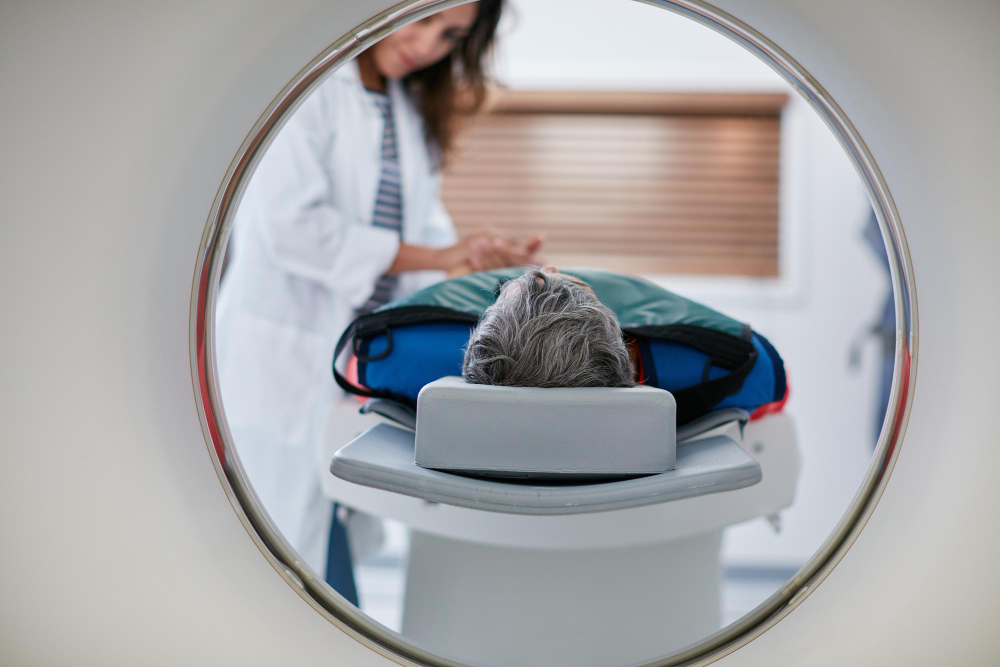
Radiation dose management in medical imaging is based on the core principles of justification and optimization, ensuring that imaging delivers the necessary diagnostic information while minimizing radiation exposure to patients. Qaelum’s DOSE, an advanced dose management system, is deployed at over 1,200 sites globally, supporting the optimization of radiation protection, imaging safety, and dose awareness in clinical practice across Europe, North America, the Middle East, Asia, and Australia. This article summarizes some examples of how DOSE has been used in clinical practice.
A German hospital network implemented many of the patient dose monitoring targets from the International Atomic Energy Agency [1] across its five sites through their DOSE installation, by analyzing radiation exposure across two CT scanners and high-volume protocols. While most scans were consistent, a notable exception occurred during a specific shift, where the computed tomography dose index (CTDI) exceeded the average by 20%: attributed to higher kVp, patient body habitus, and suboptimal arm positioning. Review of both low- and high-dose outliers highlighted the critical importance of proper patient positioning, protocol adherence, and careful selection of scan parameters, revealing optimization opportunities in reducing repeated localizers, validating scan settings, reviewing image quality in low-dose outliers, and improving data completeness [2].
Multiple sites have demonstrated the value of reviewing protocol deviations and their impact on dose. For example, analysis of four CT scanners in DOSE over one year revealed that head exams were highly standardized, with irradiation events and dose length products (DLPs) closely aligned with organizational medians, whereas thorax studies exhibited significant variability. Contrast-enhanced thorax exams further revealed inter-site differences, particularly flagging one site that was not using monitoring to trigger scans following contrast administration. Protocol inconsistencies, especially in standard and ultra-low dose thorax studies, contributed to extreme DLP outliers, underscoring the need for regular protocol review [3].
Another site utilized DOSE to compare scan settings across scanners and evaluate the effect of scan parameter deviations on organ dose. Organ doses were, on average, 57% higher in head protocols on one scanner and 76% higher in abdomen protocols on another. Certain variations in scan settings were found to increase doses to radiosensitive organs, such as the thyroid and gonads, in head and abdomen protocols, respectively [4]. Similarly, DOSE was also used to compare statistics on dose metrics, event counts, and scanned body regions. Deviations were more common on certain scanners, with repeated spiral scans found to be the primary contributing factor to higher dose [5].
High cumulative doses and dose outliers were investigated in a pediatric hospital by monitoring alerts in DOSE for cumulative doses above 50 mSv and DLP 1.3 times the American College of Radiology Diagnostic Reference Level. Few alerts were generated, indicative of good clinical practice. Analysis of these alerts identified common triggers, including repeated scans, incorrect data entry, and patient size, helping target future optimization efforts for specific exam types and age groups [6].
Image quality is equally important for protocol optimization. Global Noise Level (GNL), an automated scan-specific metric calculated in DOSE, combined with CTDI, water equivalent diameter (WED), and kVp across reconstruction methods and kernels, was collected for 46,000 exams across six scanners to benchmark quality and dose. All parameters were successfully and meaningfully integrated in a single summary plot, allowing efficient outlier detection. Findings further demonstrated the impact of kVp on image noise, reducing GNL by up to 70% when changing from 80 to 140 kVp [7].
Patient positioning is another key factor in dose optimization. Vertical positioning practices were evaluated in DOSE by quantifying their effect on radiation exposure. Offsets were found to range between -5.8 and 4.9 cm, and a negative trend was observed between patient size (WED) and vertical offset, with bigger patients positioned closer to the tube. Average tube current was 39% higher for the patient positioned with arms down compared to the patient with arms up. Tube current and CTDI were expected to increase by 6% and 19%, respectively, per 1 cm offset towards the tube [8]. Comparisons of automatic positioning cameras versus manual approaches showed significant improvements in abdomen-pelvis scans, confirming that technology can enhance positioning consistency and dose management [9].
These examples from clinical facilities worldwide have highlighted the utility of dose management, helping to ensure consistency of patient care and quality, and supporting both patient safety and clinical accuracy in imaging.
References
[1] INTERNATIONAL ATOMIC ENERGY AGENCY, Patient Radiation Exposure Monitoring in Medical Imaging, Safety Reports Series No. 112, IAEA, Vienna (2023).
[2] Romanyukha A, Heiland H, Okonkwo E, Laubenberger J, Fiebich M, Fitousi N. Determining CT dose optimization targets according to IAEA recommendations. Annual Meeting of the German Society for Medical Physics, DGMP, 24 – 27 September 2025, Berlin, Germany.
[3] Bateman L, Romanyukha A, Fitousi N. Review of study deviations in clinical practice. Managing Patient Dose - Utilising Dose Management Software, IPEM, 4 October, 2023, Manchester, UK.
[4] Romanyukha A, Solis N, Chapel M, Merino JA, Forner A, Fitousi N. Variation in scan settings within two standard protocols and its impact on organ dose. European Congress of Medical Physics, ECMP 2024, 11 – 14 September 2024, Munich, Germany.
[5] Solis N, Romanyukha A, Chapel M, Merino JA, Forner A, Fitousi N. Scanning beyond the defined protocol: how often does it happen and what is the impact on dose? European Congress of Medical Physics, ECMP 2024, 11 – 14 September2024, Munich, Germany.
[6] Romanyukha A, Fricke S, Fitousi N. Investigation of High Cumulative Doses and Dose Outliers in a Pediatric Hospital. Annual meeting of the American Association of Physicists in Medicine, AAPM 2025, 27 – 30 July 2025, Washington, DC, USA.
[7] Vignero J, Bosmans H, Petrov D, Miseur B, Binst J, Torfs K, Fitousi N. Routine Quality Monitoring in Abdominal CT Using Global Noise Reference Levels in a 4-Parameter Protocol Summary Plot. Annual meeting of the American Association of Physicists in Medicine, AAPM 2025, 27 – 30 July 2025, Washington, DC, USA.
[8] Romanyukha A, Fitousi N, Dalah E. Mispositioning practices and their effect on radiation exposure in abdomen CT exams. European Congress of Radiology, ECR 2024, 28 February- 3 March 2024, Vienna, Austria.
[9] Romanyukha A, Dalah E, Fitousi N. Comparison of vertical positioning offsets in chest and abdomen scans between automated and manual patient positioning. American Association of Physicists in Medicine Annual Meeting, AAPM 2024, 21 – 25 July 2024, Los Angeles, USA.
Solutions