Risk Communication in Medical Imaging: Key Issues and Current Approaches
June 11, 2026
Anna Romanyukha, PhD; Niki Fitousi, PhD
Risk in medical imaging, particularly the balance between diagnostic benefit and potential harm from ionizing radiation, has long been a topic of scientific discussion. Recent media coverage of the potential radiation induced cancer risks due to medical exposure has refocused the scientific community on the challenge of meaningfully assessing and communicating risk.
This topic was addressed in a physics session at the 2025 Annual Meeting of the Radiological Society of North America, titled “Un”risky Business: Easing the Uncomfortable Relationship Between Risk and Benefit in Medical Imaging [1]. In this session, total risk was defined as the sum of clinical risk and radiation risk, emphasizing that risk assessment cannot be based on radiation exposure alone. Clinical risk is often a qualitative measure, defined as misdiagnosis and often based on the radiologist’s subjective opinion on the image quality. To address this limitation, a mathematical model of clinical risk was presented, incorporating quantitative parameters including disease prevalence within the imaged population, differences in survival associated with correct versus incorrect diagnoses, life expectancy loss resulting from misdiagnosis, and the ratio of false-positive to true-positive outcomes. The latter was associated with the computed tomography (CT) dose index or CTDIvol, i.e., the concept that higher dose results in better image quality. Radiation risk was defined using a risk index according to the United States National Research Council’s Committee to Assess Health Risks from Exposure to Low Levels of Ionizing Radiation (BEIR VII) framework, which offers patient age- and sex-specific lifetime attributable risk (LAR) estimation methodology [2]. Radiation risk also incorporated disease prevalence and false-positive to true-positive ratios [3].
The combined model was evaluated on a cohort of one million digital twins simulating a liver cancer scenario, with simulations based on survival data from the U.S. National Cancer Institute. Results demonstrated that clinical risk, expressed as mortality per 100 patients due to misdiagnosis, was approximately five times greater than radiation risk. This imbalance was further highlighted through comparisons of mortality risks associated with different aspects of cancer care. While radiation-induced cancer risks are frequently discussed, mortality associated with surgical intervention and chemotherapy is substantially higher, with reported rates of approximately 1.32% for surgery compared with 0.04% for radiation-induced malignancy [3]. Despite this disparity, radiation risk continues to dominate both scientific discourse and public perception, often without adequate contextualization.
Speakers in the session emphasized that effective risk management in medical imaging requires rigorous dose and image quality optimization, guided by principles that are task-, patient-, procedure-, and equipment-specific [4]. Finally, the importance of clear and balanced communication was stressed, underscoring that patient communication should be sensitive, informed, and engaging [5].
In the context of dose management, radiation risk remains the most relevant measure for guiding optimization efforts and communication strategies. In response to customer demand, a LAR calculation was recently implemented within Qaelum’s dose management system, DOSE. This implementation follows the International Commission on Radiological Protection (ICRP) methodology for LAR estimation, assuming a dose and dose-rate effectiveness factor (DDREF) of 2 for solid cancers and applying a Euro-American composite population model [6].
LAR calculations are conventionally based on organ-specific doses and LAR coefficients, but effective dose offers a simplified, alternative approach that has been employed previously [7-9]. To evaluate the comparability of these approaches, LAR estimates based on organ dose (LAROrganDose) and effective dose (LAREfDose) were assessed in a cohort of 19,248 spiral CT acquisitions of standard anatomical regions (abdomen, chest, and head) [10]. Across all examination types, LAREfDose tended to yield higher values than LAROrganDose, with the largest discrepancies observed for head acquisitions. Both effective dose and corresponding LAR values were 2-13 times lower for head CT compared with chest and abdominal CT.
Differences between LAR estimation methods were more pronounced in younger patients, consistent with age-dependent risk coefficients. The median difference between LAROrganDose and LAREfDose was -0.006 cases per 100 patients for those under 50 years of age, compared with -0.001 cases per 100 patients for those aged 50 years and older. Differences were largely attributable to dose calculation assumptions: effective dose is derived from dose-length product (DLP) and therefore accounts for scan length, whereas organ dose calculations rely on CTDIvol and assume standardized scan ranges for a given anatomical region (Figure 1). Consequently, examinations with extended scan lengths produced larger discrepancies between the two approaches. Overall, differences in LAR estimates ranged from 0.001 to 0.003 cases per 100 patients, indicating that both methods provide broadly comparable estimates, with the effective-dose-based approach yielding a more conservative risk assessment.
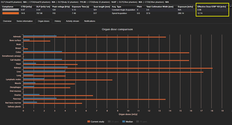
Figure 1. Effective dose (top) and organ doses (bottom) as displayed for an exam in DOSE.
Although such radiation risk estimates are derived from population-based epidemiological models and are not intended for application at the individual patient level, there remains a growing need within the scientific and clinical communities for risk metrics that extend beyond traditional dosimetric quantities and can be understood by non-experts. When implemented within automated dose management systems, such metrics can provide consistent indicators of radiation risk. Importantly, the interpretation and application of radiation risk metrics should be situated within a broader clinical risk-benefit framework that accounts for the often substantially greater risks associated with misdiagnosis and therapeutic interventions, ensuring that radiation risk is neither underestimated nor disproportionately weighted in clinical decision-making.
For more information about Qaelum’s DOSE dose management system, please visit: https://qaelum.com/solutions/dose
References
[1] Samei E. S5-CPH14AAPM/RSNA Physics Tutorial 2: “Un”risky business: Easing the uncomfortable relationship between risk and benefit in medical imaging. Presented at: Radiological Society of North America Annual Meeting (RSNA 2025); November 30, 2025; Chicago, IL. Available at: https://cattendee.abstractsonline.com/meeting/21232/Session/493
[2] National Research Council (NRC) Committee to Assess Health Risks from Exposure to Low Levels of Ionizing Radiation. Health Risks from Exposure to Low Levels of Ionizing Radiation: BEIR VII Phase 2. National Academy of Sciences; Washington, DC: 2005.
[3] Ria F. Theoretical Approach to Risk/Benefit Assessment. Radiological Society of North America Annual Meeting, RSNA 2025, November 30-December 4, 2025; Chicago, IL.
[4] Trianni A. From a Physicist Perspective. Radiological Society of North America Annual Meeting, RSNA 2025, November 30-December 4, 2025; Chicago, IL.
[5] Frush D. From a Radiologist Perspective. Radiological Society of North America Annual Meeting, RSNA 2025, November 30-December 4, 2025; Chicago, IL.
[6] ICRP 147: ICRP, 2021. Use of dose quantities in radiological protection. ICRP Publication 147. Ann. ICRP 50(1).
[7] Hanley M, Koonce J, Bradshaw M. X-ray Risk. Accessed February 5, 2025. https://www.xrayrisk.com/.
[8] Wurster, C.D., Winter, B., Wollinsky, K. et al. Intrathecal administration of nusinersen in adolescent and adult SMA type 2 and 3 patients. J Neurol 266, 183–194 (2019). https://doi.org/10.1007/s00415-018-9124-0
[9] Aw-Zoretic J, Seth D, Katzman G, Sammet S. Estimation of effective dose and lifetime attributable risk from multiple head CT scans in ventriculoperitoneal shunted children. Eur J Radiol. 2014 Oct;83(10):1920-4. doi: 10.1016/j.ejrad.2014.07.006. Epub 2014 Jul 16. PMID: 25130177; PMCID: PMC4623705.
[10] Romanyukha A, Vignero J, Bosmans H, Fitousi N. Comparison of radiation risk assessment methodologies for standard CT exams. European Congress of Radiology, ECR 2025, 26 February- 2 March 2025, Vienna, Austria.
Authors
Anna Romanyukha received her Ph.D. degree in medical physics from the Centre of Medical Radiation Physics (UOW, Australia) and her M.Sc. degree in health physics from Georgetown University (Washington DC, USA). She worked as a post baccalaureate and pre doctoral fellow at the National Cancer Institute (NIH, Washington DC) on various projects including radiation dose estimation from diagnostic exposures. She now works in Qaelum NV, focusing on advanced software tools in patient radiation dose management and quality.
Niki Fitousi, PhD, is a certified medical physicist with professional experience in all fields of Medical Physics (Radiation Therapy, Diagnostic Radiology, Nuclear Medicine, Radiation Protection). She is currently the Head of Research and Development in Qaelum, focusing mostly in the fields of radiation dose management, quality and efficiency in medical imaging. She is also a member of the Medical Physics World Board of the International Organization for Medical Physics, as well as other Medical Physics organizations.
Solutions